Peering into the unseen world, microscopes have revolutionized our understanding of everything from cells to crystals. But what’s beneath the eyepiece? How does a simple arrangement of lenses and light transform the invisible into the visible?
In this article, we’ll explore the intricate inner workings of a microscope, guided by the detailed diagram, for smoothly transitioning between concepts. We’ll demystify the complex components and explain how they interact to magnify the minuscule. So, whether you’re a science student, a curious hobbyist, or a seasoned researcher, let’s dive into the fascinating world of microscopes together.
Understanding the Diagram Microscope

Diving deeper, let’s unlock the enigma of the Diagram microscope, interpreting its unique design and inherent intricacies. Key elements that stand out set this microscope apart, providing powerful investigative capabilities. Maximizing Your Wins with this instrument requires understanding these elements.
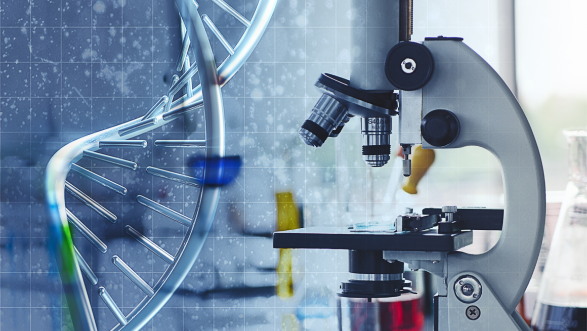
Examination of the Diagram microscope reveals a meticulously designed configuration. Boasting a solid, stable base, and an arm extending vertically, it provides support to the core components. The stage, positioned midway on the arm, serves as a platform for the specimens being observed. The revolving nosepiece, located above the stage, holds the microscope’s multiple objective lenses. Each lens represents different magnification powers, presenting a range for varied inspection needs. For instance, a 4x objective provides low magnification for viewing larger specimen areas, while a 100x oil immersion lens offers high magnification for detailed cellular examination.
Navigating the Diagram Microscope

Transitioning from understanding the components of the Diagram microscope, it’s time now to navigate using a step-by-step interpretation and understanding of common symbols for proficiency.
Current Updates and Enhancements
Overcoming previous limitations, present-day Diagram microscopes exhibit considerable improvements. One notable uplift concerns resolution quality. Modern revisions present sharper, better-focused images, thanks to advanced lens technology. This factor drastically increases the microscope’s efficacy in discerning otherwise indistinguishable features.
Lighting issues no longer pose a viewing obstacle, courtesy of modern illumination techniques that perfect visualization. The addition of adjustable brightness ensures users can tailor viewing conditions to better suit their needs.
Arguably most significant is the integration of digital technology. This facilitates seamless data collection, processing, and sharing. Users can digitally annotate structures, significantly reducing manual effort and increasing efficiency. The microscope’s design has also been streamlined for better ergonomics, making it easier to transport and use.
All these updates and enhancements anchor the Diagram microscope’s relevance and usability in today’s scientific and educational fields, powering minute explorations that deepen the understanding of our world.
Comparing the Diagram Microscope with Other Microscope Diagrams

In contrast to other microscope diagrams, the Diagram microscope serves a unique role, demonstrated by its exceptional features, extensive usability, and potential areas for growth.
Advantages of the Diagram Microscope
Compared to other microscope diagrams, it features superior resolution quality and digital integration capabilities. Central to this is its extraordinary resolve for minute details, empowering researchers to observe intricate structures in bacteria, cells, and genes. This clarity of vision enables swift and accurate diagnosis in the medical field, making it an indispensable tool in disease research.
Further, its digital integration outperforms that of competing microscopes. Diagram microscope is seamlessly compatible with latest software, thereby simplifying both the data collection process and the sharing of findings across various platforms. It sparks interactive learning in educational contexts, making complex biological concepts tangible for students.
Potential Drawbacks and Improvements
Despite its remarkable capabilities, Diagram microscope isn’t flawless. Initial iterations were marked by problems such as subpar image clarity and lackluster digital integration. However, these issues were largely tackled in the modern versions, indicating a trend towards continuous improvement.
There’s still room for advancement, nonetheless. One key area is the user interface which, although improved, could further enhance user experience by being more intuitive and responsive. Further, increased versatility in image magnification options could augment the viewing experience, especially when dealing with highly minute or extensive sample areas. As it adjusts to advancements, the Diagram microscope has a potential for setting further milestones in microscopy.


